Foot surgery refers to the orthopaedics specialisation that treats some of the most common foot pathologies including hallux valgus and hallux rigidus, distal valgus of the big toe, metatarsalgia, hammer toe, “claw” toe, varus of the 5th toe and Morton’s Neuroma.
Foot surgical interventions rely both on traditional surgical techniques as well as minimally invasive ones. The latter make it possible to avoid traditional surgical “cuts”, using very small burrs with are introduced through small skin incisions. These small burrs model the bone and make it possible to correct the deformity.
The most significant causes of deformities of the foot are attributable to two major syndromes, deriving from an incongruous support of the foot: pronatory syndrome and supinatory syndrome.
Pronatory syndrome
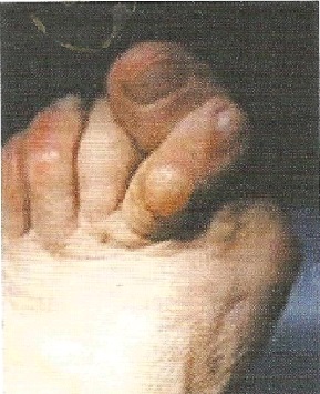
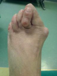
The foot rolls towards the inside, while the big toe is often osteoarthritic and turned outwards. The toes are dislocated on the metatarsophalangeal joints and assumes a hammer like position. Often, if left untreated, the second toe not only flexes, but overlaps the big toe. This syndrome is always associated with a metatarsalgia, namely painful plantar calluses, which in many cases are the driving reason that cause the patient to consult a specialist.
Supinatory syndrome
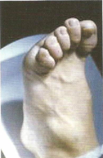
Supinatory syndrome is characterised by an exaggerated dorsiflexion of the toes, due to the prevalence of the contraction of the extensor tendons. The toes take on a “claw” like appearance.
Both cases imply the presence of a hallux valgus, condition characterised by the deviation of the big toe towards the outside. When this happens, the full extensor apparatus shifts laterally and the first metatarsal angles medially. These changes in the anatomy of the foot tend to become chronic, worsening the hallux valgus of the big toe. As a result, an exostosis of the head of the first metatarsal bone forms, characterised by a medial bone protrusion with redness of the skin, the lateral displacement of the first phalanges and a consumption of articular cartilage, leading to the evolution of the condition into osteoarthritis.
Metatarsalgia: treatment of the condition
 When the condition is at an advanced stage, the other toes assume a hammer like position and painful callouses appear under the sole of the foot (metatarsalgia).
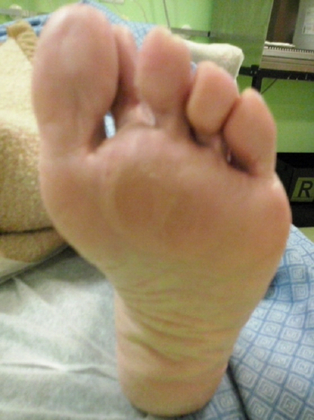
Metatarsalgia is a localised pain in the anterior region of the sole of the foot, which appears or is accentuated when weight is placed on the foot. The pain may be associated with the presence of calluses and a “claw” like deformity of the toes. Under normal conditions, there is an even distribution of the weight placed on the forefoot, supported by the correct metatarsal alignment on the front and dorso-plantar planes. In the event of disharmony in the length of one or more metatarsals and of anomalies in the inclination of the back of the foot or the forefoot, the correct distribution of weight on the foot is compromised. In more acute stages the pain can make it difficult to walk, which in some cases leads to abnormal foot support positions, resulting in disorders of the knee and spine.
The causes may be structural, congenital, traumatic and/or inflammatory in nature. One or more metatarsals longer than the standard or tilted downwards will cause an overload. Conversely, shorter metatarsals sloping upwards will be insufficient to support the weigh, overloading those next to them, in both cases resulting in a metatarsalgia.
In mild and reducible forms, the biomechanical alteration can be corrected through the use of orthotics, while the pain can be treated using medical or physical therapy. In structured forms, which are no longer correctable, a surgical intervention is necessary to restore the correct metatarsal realignment.
The metatarsal intervention is performed using a minimally invasive percutaneous technique or a traditional technique through a small surgical incision (neither technique makes it possible to repair the joint in such a way as to allow the patient to regain a normal walk).
Percutaneous surgery makes it possible to treat the metatarsalgia using two or three small incisions on the back of the foot that allow the surgeon to intervene using a mico burr on the neck of the painful heads. The osteotomies produced are not fixed through means of synthesis but left free. The use of rigid shoes and immediate weight on the foot facilitate the correct repositioning.
Hammer toe: treatment of the condition
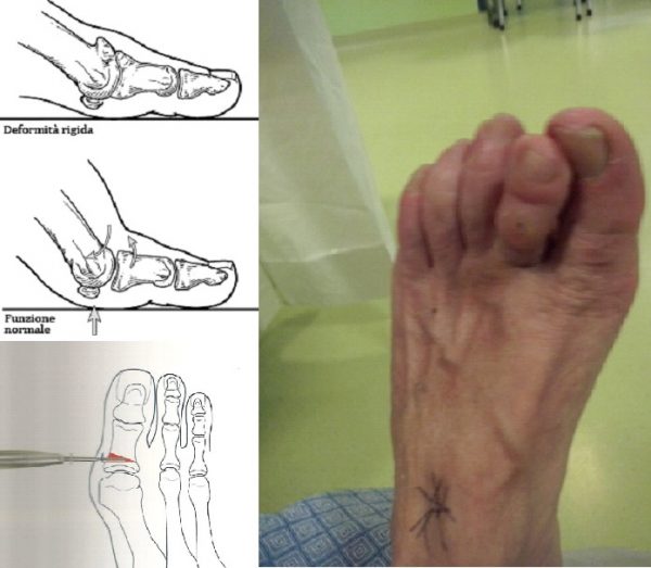
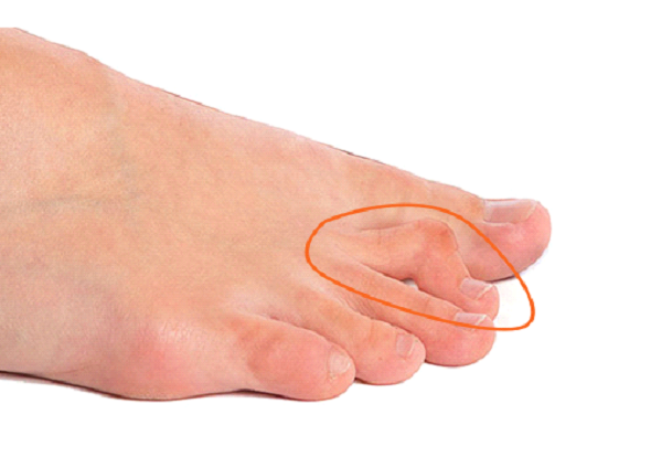 Hammer toe is a deformation characterised by flexion of the proximal interphalangeal joint, which causes the toe to assume the shape of a piano key gavel. In addition to the aesthetic factor, this condition causes pain that hinders the use of footwear. The surgical intervention to return the full function of the toe is carried out in a Day Hospital with a simple tenotomy of the flexors and of the extensors at the base of the phalanges, performed percutaneously or alternatively through a small surgical incision, with osteotomy of the proximal phalanges, or, in severe cases associated with lateral or medial deviation of the toe, with arthrodesis of the interphalangeal joint.
Heel spur: treatment of the condition
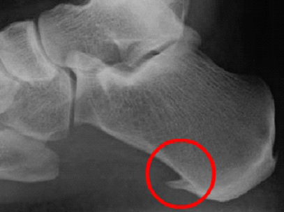
Heel spur or plantar spur consists of an excessive growth of the heel bone and occurs when the plantar fascia applies excessive traction on the heel. Most cases can be treated with shock wave “bombardment”, which disrupt the areas presenting calcareous infiltration. If the condition persists, a surgical intervention can be carried out through a small incision along the edge of the heel to remove the spurs.
Haglund's disease: treatment of the condition
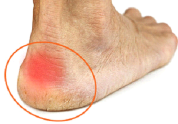
Haglund’s disease is a painful inflammation of the heel, often caused by varus of the foot. This condition can be treated using innovative anti-shock insoles, which avoid overloading the joints. If the condition persists, a surgical intervention can be carried endoscopically or alternatively through a small surgical incision and consists in the resection of the posterior-superior angle of the heel. The patient can immediately put weight on the foot.
Morton's Neuroma: treatment of the condition
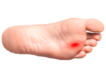
Morton’s neuroma is a condition affecting the interdigital nerve responsible for the sensitivity of the internal part of two adjacent toes.
The condition is characterised by an enlargement of the nerve (neuroma) or by a secondary compression of the same by adjacent structures, at the level of the metatarsal heads.
Both these phenomena give rise to the typical pain and paraesthesia (tingling, tremors, pins) which tends to radiate through the two innervated toes when the foot rests on the ground during walking.
Excision surgery for treating Morton’s neuroma is performed through a small dorsal incision.
Flat foot: treatment of the condition
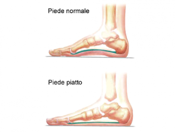
Pronatory or flat foot syndrome is a condition characterised by a lowering of the inner plantar arch of the foot, that can even reach the extreme limit of becoming completely flattened, resulting in degeneration of the tendon and the onset of medial or lateral pain (valgus foot). When, around puberty (from 10 to 14 years of age), the foot has not fully matured and a significant flat-footedness persists, it is possible to intervene with minor surgery, achieving the immediate correction of the arch.
Flat foot surgery is a simple and minimally invasive procedure consisting of inserting a screw into the calcaneus at the tarsal sinus level (empty space in front of the external malleolus) through a small skin incision. The child is discharged from the clinic on the same day as the surgery. The next day, the child can already support the foot and after approximately a week, they are able to walk without support.
Hallux valgus: treatment of the condition
Various surgical techniques are used to correct hallux valgus.
The hallux valgus intervention is performed using a minimally invasive percutaneous technique or a traditional technique through a small surgical incision (neither technique makes it possible to repair the joint in such a way as to allow the patient to regain a normal walk).
The percutaneous technique is the most advanced surgical procedure currently available to treat hallux valgus, making it possible to intervene with just two 2 mm incisions through which the deformed bones are modelled, obtaining good results and facilitating a much faster recovery compared to traditional techniques.
Hallux rigidus: treatment of the condition
In many cases in which the load of the first ray of the foot is altered, a common condition develops known as hallux rigidus. The condition relates to a primary or secondary osteoarthritis of the metatarsal phalangeal joint of the first toe. The condition is characterised by limited mobility of the joint associated with pain and often to the presence of osteophytes (bony growths) on the dorsum of the foot, between the head of the first metatarsal and the base of the proximal phalanges, making it difficult to extend the big toe while walking. Surgical correction of hallux rigidus facilitates the surgical excision of the osteophytes and the remodeling of the joint. The hallux rigidus intervention is performed using a minimally invasive percutaneous technique or a traditional technique through a small surgical incision (neither technique makes it possible to repair the joint in such a way as to allow the patient to regain of a normal walk). In the case of hallux rigidus, it is often only the distal phalanges of the big toe that deviates outward. This condition is known as distal valgus of the big toe and is easily treatable using a small wedge shaped osteotomy of the basal phalanges of the big toe.
Ankle instability: treatment of the condition
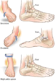
Ankle instability is a condition characterised by frequent collapse of the ankle associated with a persistent swelling primarily localised on the outside of the ankle. The patient will also experience pain when walking, especially on uneven ground.
The most frequent cause is represented by one or more distortions leading to a rupture of the external ligaments or pathological changes inside the joint.
In case of non-treatment, the injured ligaments do not regain the tension they initially had before the trauma and the joint remains exposed to other possible distortions. Surgical treatment of the condition consists of the repair of an injured ligament through a small skin incision along the side of the ankle. If the ligaments are damaged to the extent that they cannot be directly repaired, healthy tissue is taken from the area surrounding the lesion and transplanted in place of the damaged ligaments. After the surgical procedure the ankle is immobilised for four weeks, followed by a subsequent rehabilitation therapy.
Ankle surgery: arthroscopy of the ankle
Arthroscopy of the ankle is a type of minimally invasive surgery, performed using a fibre-optic scope. By observing the ankle joint a simple incision can be made that makes it possible to intervene on the damaged part. This procedure avoids large incisions and is able to treat a variety of joint conditions.