Hand surgery currently necessitates a multidisciplinary preparation involving orthopaedics, traumatology, plastic surgery and reconstructive microsurgery, in addition to basic general surgery preparation. This multi-specialist training allows specialists in this field to better treat inflammatory and degenerative conditions, traumatic lesions and their debilitating outcomes, as well as congenital malformations. Surgical procedures involving the hand are now able to treat or at least improve any condition concerning the specific area, restoring the injured patient’s hand grip function. After excluding other alternative treatments, the hand surgeon will select the surgical procedure deemed necessary to treat the patient.
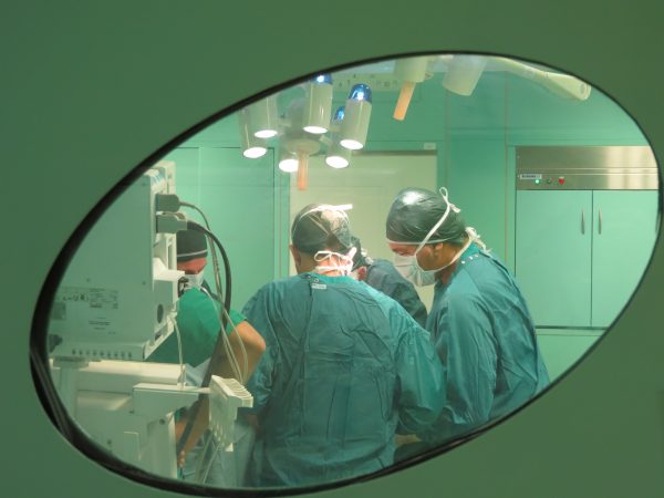
The Casa di Cura San Giovanni clinic has organised a team of expert surgeons specialised in orthopaedics and plastic surgery who have at their disposal dedicated clinics, new high-tech operating rooms at the forefront in terms of all sterilisation and safety protocols, the very best diagnostic tools available for this type of surgery and an analytical laboratory open daily which performs exams at any time and as needed.
Our specialists are able to perform any type of surgical procedures involving the hand. Our hand surgery centre stands out for the professionalism and technical skills offered. Each year, our surgeons perform more than 800 surgeries to treat hand and wrist conditions. An overview of the most common conditions treated by hand specialists who work at our facility are presented below:
Trigger finger: surgical intervention and treatment of the condition
Stenosing tenosynovitis of the finger flexors (also known as trigger finger or trigger thumb) most commonly affects the flexor tendons of the fingers.
The condition is characterised by a thickening of the tendon sheath that covers the flexor tendons and facilitates, in normal conditions, the sliding movement in the osteofibrose structures present in the palm of the hand and in the fingers. These osteofibrose structures called pulleys, keep the tendons attached to the deep planes during the contraction of muscles and prevent dislocation.
When a hypertrophy of the membrane lining the tendons or an increase in the rigidity of these structures manifests itself, the normal sliding movement of the tendons can be compromised, resulting in the appearance of “trigger” sensations during finger movements or incomplete extension of the corresponding finger. Stenosing tenosynovitis is often caused by inflammations, micro-traumatic stress or hereditary factors and initially manifests itself with pain in the distal palmar region (at the base of the fingers) during the flexion-extension of the affected finger, then subsequently with the onset of the “trigger” effect and difficulty extending the finger, and finally with blocked flexion and limited extension of the finger. The typical surgical procedure to treat the condition is performed under local anaesthesia through an incision at the base of the finger that frees the tendon by cutting the pulley.
De Quervain’s disease treatment
De Quervain syndrome is a stenosing tenosynovitis (acute or chronic inflammation) of the short extensor tendons and the long thumb abductor.
The condition is characterised by difficulties with the long abductor tendon and the short extensor of the thumb sliding within the common osteofibrous channel that contains them, along the radial edge of the wrist, as well as by inflammation, rheumatic episodes and post-traumatic episodes. Initially this condition manifests itself with pain experienced during flexion and extension of the thumb. The pain tends to increase when, with the thumb within the palm of the same hand, the patient is encouraged to perform a forced movement characterised by the ulnar deviation of the wrist.
De Quervain’s disease involves progressive difficulty using the thumb as a result of acute pain associated with its movement and the progressive emergence of serious functional impairment of the thumb and consequently of the hand, with the onset of permanent pain. The surgeon’s goal is to relieve the pain through a surgical intervention performed under local anaesthesia using a mini hand incision that eliminates the friction between the sheath and the tendons responsible for the inflammation and pain experienced by the patient.
Dupuytren's disease: surgical intervention and treatment of the condition
Dupuytren’s disease is a condition characterised by the thickening and subsequent retraction of the palmar aponeurosis, a typically thin subcutaneous hand membrane located in the palm of the hand and the fingers.
During the progression of the condition, this membrane thickens, initially resulting in localised nodular formations and subsequently in chord-like formations in the palm of the hand or extending up to the fingers, with the gradual retraction of the palm and flexion of the fingers. There are several pathogenetic hypotheses relating to the condition. However, the actual cause of the advancement of the disease, with gradual and progressive flexion deformity of the fingers and consequent functional limitation is not yet well understood.
The gradual flexion of the fingers induced by the progression of the condition may result in skin retraction and joint alterations, with consequent secondary deformity resulting in ankylosis or subluxation or joint dislocation.
The surgical intervention to correct problems arising from Dupuytren’s disease is carried out using a small needle. The procedure is painless, it does not leave marks or scars and does not require long periods of rehabilitation. In more severe cases the surgeon may resort to an aponeurotomy of the palm and fingers.
Rhizarthrosis: surgical intervention and treatment of the condition
The trapeziometacarpal joint, located at the base of the first ray of the hand, facilitates the ab-adduction and back movement of the thumb. The saddle joint conformation facilitates a broad mobility of the thumb, at times at the price of significant instability with secondary osteoarthritic degeneration due to hyper-use.
Idiopathic osteoarthritis disease, traumatic joint lesions, rheumatoid arthritis or other connective tissue conditions often cause destructive alterations of the trapeziometacarpal joint. The clinical manifestations of the condition are more frequent in perimenopausal females. In the early stages of the disease, the treatment may consist of infiltration of hyaluronic acid or minimally invasive outpatient procedures which facilitate an almost immediate recovery. When osteoarthritis degeneration irreversibly alters the ability to maintain good joint movements and leads to pain that cannot be controlled using normal physical-rehabilitative and/or drug therapies, a surgical intervention is recommended.
In the past, the surgical treatment for rhizarthrosis was arthrodesis of the trapeziometacarpal, with consequent loss of movement of this joint. Modern orthopaedic treatment of a symptomatic osteoarthritis joint aim to restore a complete and painless movement thereof. Over the last decade the most frequent surgical treatment was the removal of the trapeze with tendon interposition and inevitable shortening of the first ray of the hand.
Current spacer prosthesis overcame the inconveniences of the first prosthesis to the bone anchorage and their favourable evolution has facilitated a painless recovery of the movement of the base of the thumb, with good grip strength. The possible need to remove the prosthesis inserted, leads back to the established effectiveness of an arthroplasty (trapeziectomy and tendon interposition).
In some cases treatment with the partial or total excision of the trapezium, without the introduction of the prosthesis may still be relevant.
Carpal tunnel syndrome: surgical intervention and treatment of the condition
Carpal tunnel syndrome is a very common condition especially in females, caused by compression of the median nerve in the carpal tunnel of the wrist. The carpal tunnel is made up of the bones of the carpus (carpal tunnel floor) and the transverse ligament (ceiling of the carpal tunnel).
Carpal tunnel syndrome is caused by micro-traumatic, inflammatory or post-traumatic factors (outcomes of wrist fractures). The symptoms experienced by the patient include: paresthesia (tingling) and sometimes pain especially at night and initially localised to the thumb, the index, the middle and the ring finger. This symptomatology is the result of irritation of the median nerve. Subsequently, with the progress of the lesion, such conditions are perceived at the level of the forearm and of the arm and may be associated with loss of tactile skin sensitivity of the hands and the loss of strength, with the patient frequently dropping objects. In the presence of such symptoms, it is essential to perform an electromyography which allows the specialist to make a precise diagnosis in relation to the area where the median nerve is compressed and the amount of damage suffered by the nerve fibres that make up the median nerve.
Initial treatment is based on functional rest with local or generally administered anti-inflammatories, together with the use of wrist orthosis and neurotrophic factors. In more advanced stages or after the failure of the therapies described, a surgical intervention is recommended. The intervention consists in the opening of the carpal tunnel and the freeing of the median nerve.